
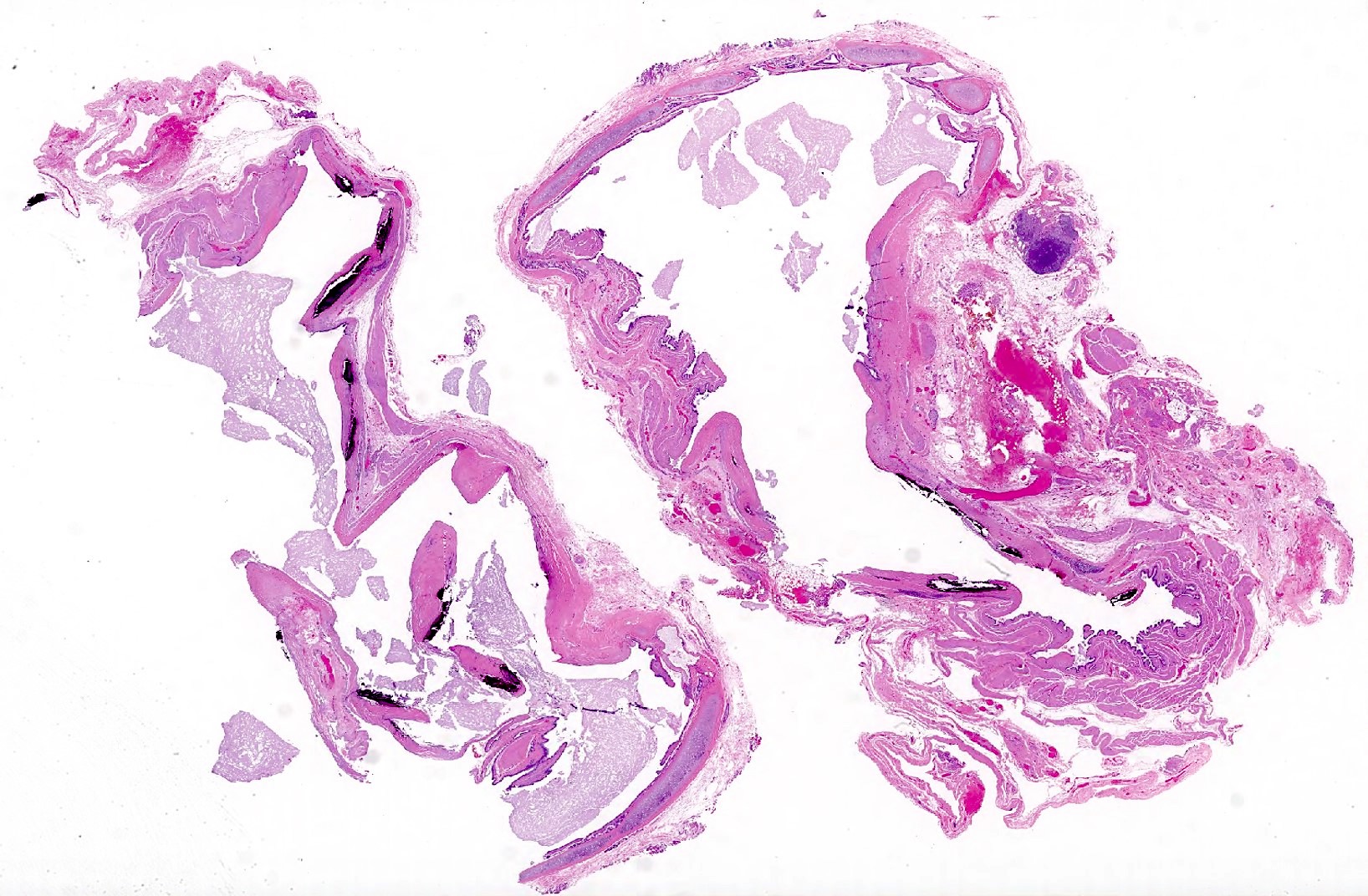
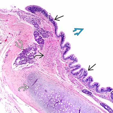
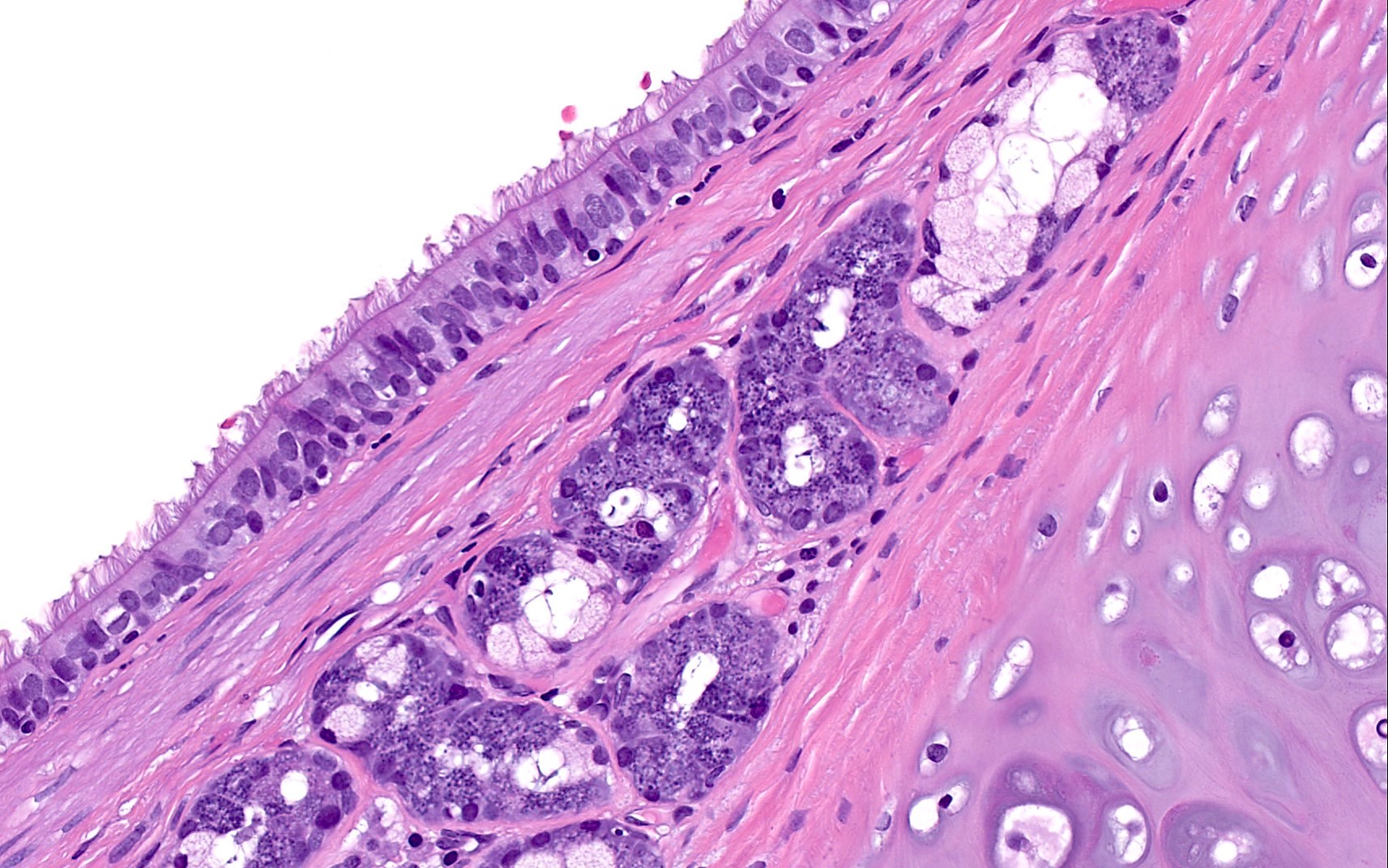
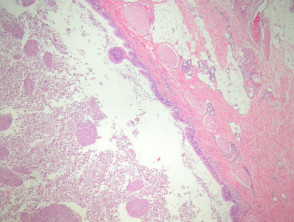
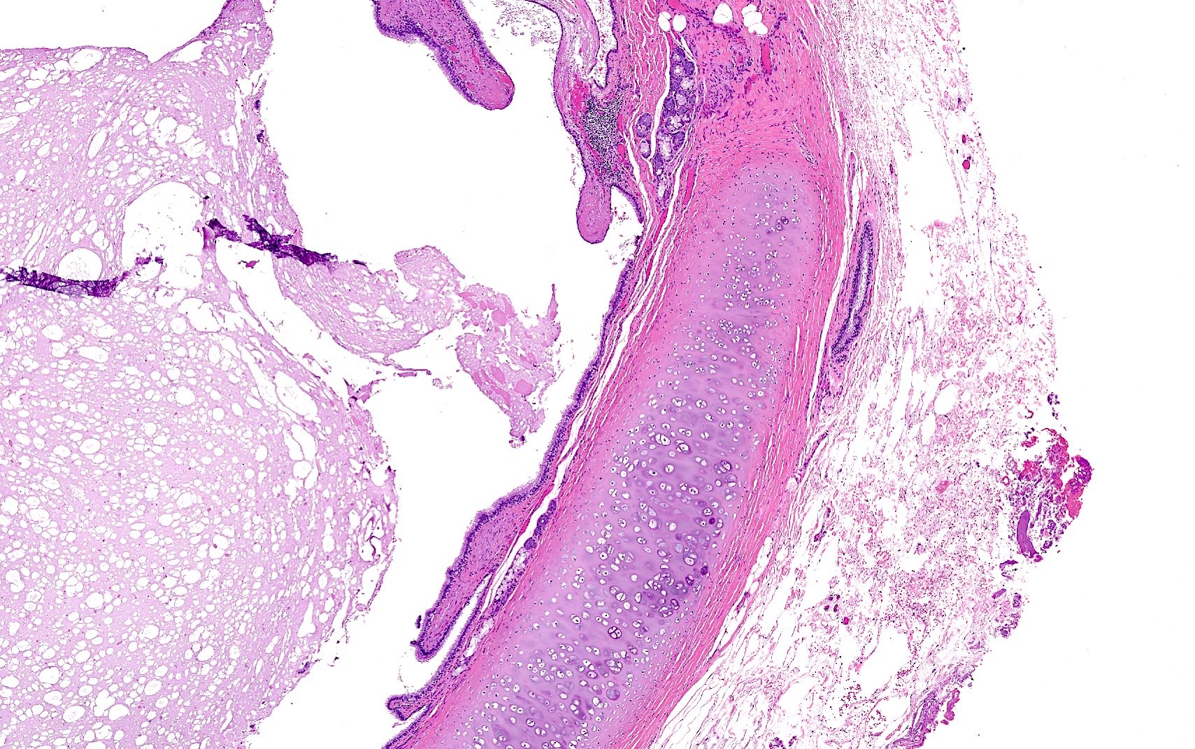
Bronchogenic cysts are mostly benign congenital abnormalities originating from the remnants of the primitive foregut. 1 they typically consist of a thin fibrous capsule lined by secretory respiratory epithelium columnar or cuboidal ciliated epithelium and may contain cartilage glandular tissue and smooth muscle.
Pathology Outlines Bronchogenic Cyst
To characterize the imaging features of bronchogenic cysts.
. In addition the study analyzed the existing literature on these lesions. The presence of this tumor on the retroperitoneum is extremely rare and high suspicion is paramount to reach preoperative diagnosis. We present here a case report of a retroperitoneal bronchogenic cyst.
The computed tomographic CT andor magnetic resonance MR or ultrasonographic images in 68 histopathologically proved cases of bronchogenic cyst in 38 male and 30 female patients aged newborn to 72 years mean 22 years were retrospectively. They are located close to the trachea or main stem bronchi. In this series however 40 per cent of the cysts were intrapulmonary Bronchogenic cysts are true cysts since they contain fluid and have an epithelial lining.
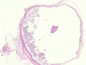
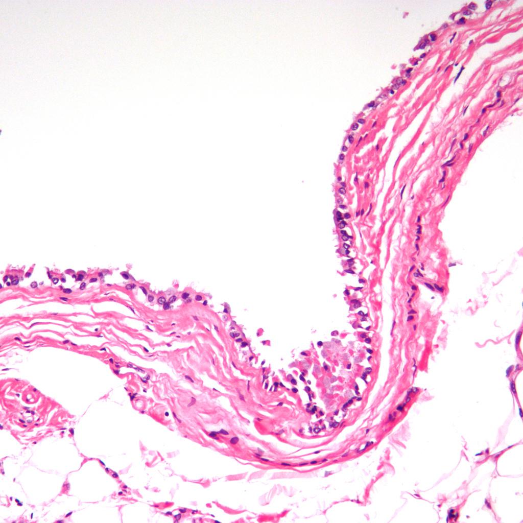
This benign cyst usually appears as a solid mass on the x. Unilocular cyst lined by respiratory type epithelium. On T1-weighted images lesions may vary in their intensity because of their protein content.
Excellent prognosis with complete surgical excision. Histology of bronchogenic cyst. Bronchogenic cysts can be intra- or extrathoracic.
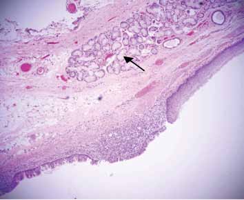
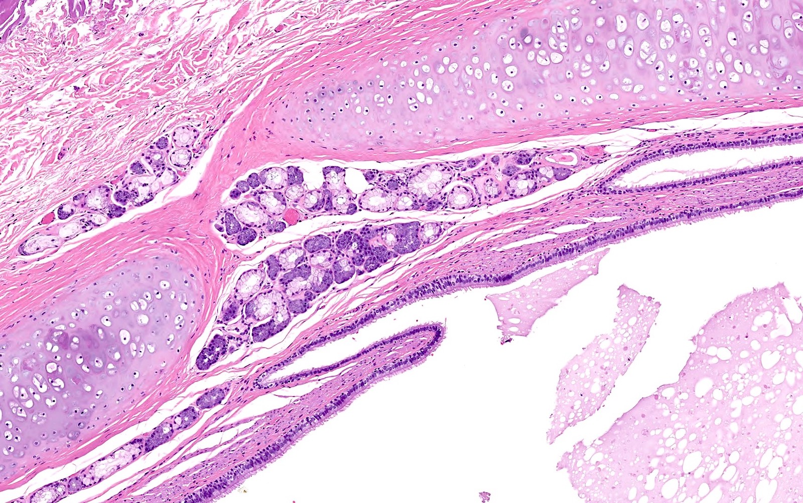
A retroperitoneal location is rare. Bronchogenic cysts are foregut-derived cystic malformations of the respiratory tract. Mucinous glands smooth muscle and sometimes cartilage are found in the wall or near the cyst.
Bronchogenic cysts are thought to result from abnormal budding of the developing tracheobronchial tree with separation of the buds from the normal airways. A study of preoperative MDCT concluded that axial MDCT. One mediastinal bronchogenic cyst 2018 S.
Histologically these are also composed of cartilage smooth muscle fibrous tissue and mucous glands. The majority of cases reported in the literature have been found in the paediatric population few cases in adults. We present a case of an upper midline pre-sternal abscess in a six-year-old female.
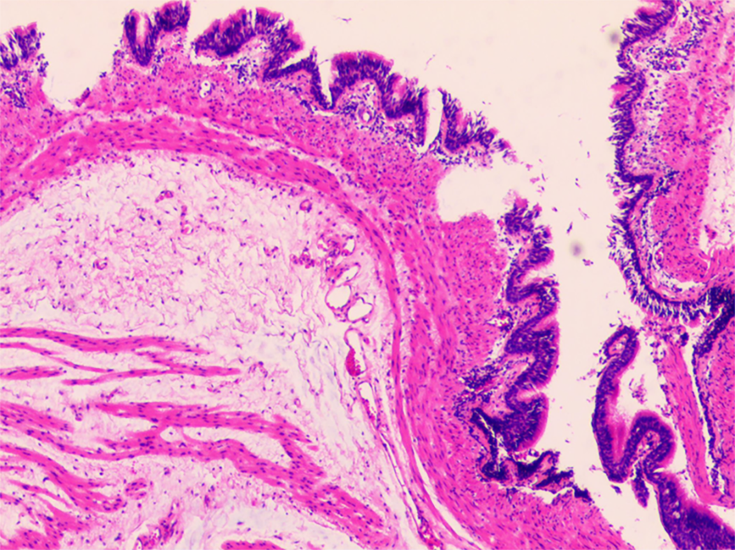
In all other locations diagnosis cannot be as reliably forecast. The lungs and pleural spaces are clear. Bronchogenic cysts may be unilocular figure 1 or multilocular.
They are usually located within the mediastinum at an early stage of gestation or in the lung at a later stage. Occurs in mediastinum lung head and neck skin abdomen retroperitoneum. The heart size is normal.
They should therefore be differentiated from non-epithelialized cystic lesions. Case Discussion The lesion was resected. However their location can be anywhere along the.
They may be located in the dermis or subcutis and occasionally drain into the overlying epidermis via a sinus. The finding of a cystic lesion at the level of the carina on CT scan or MRI is most frequently associated with a bronchogenic cyst. Due to the mostly asymptomatic behavior and the historical confusion regarding histology an exact prevalence is not known.
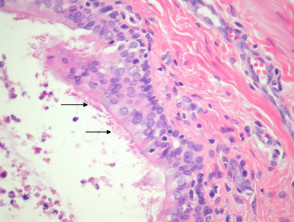
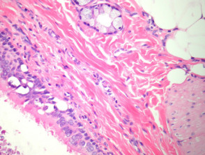
1 article features images from this case 24 public playlist include this case. They are lined by respiratory type ciliated epithelium which is characterized by cilia. Bronchogenic cysts are usually solitary asymptomatic mediastinal masses which may present at any age.
Being a rare condition bronchogenic. Histopathology Bronchogenic cysts are usually lined by ciliated columnar epithelium of respiratory type leading to distention as a. They may occur in every location along the embryonic foregut and arise from an abnormal branching of the tracheobronchial tube 12.
Pathology Bronchogenic cysts are formed in the 6th week of gestation from an abnormal budding of the tracheal diverticulum. Gastric bronchogenic cysts are rare lesions first described in 1956 with only 34 cases reported in the literature to date. Bronchogenic cysts originate from the ventral foregut that forms the respiratory system.
Cyst wall recapitulates bronchial wall with variable amounts of seromucinous glands cartilage and smooth muscle. A good majority of bronchogenic cysts 65 to 90 are mediastinal. Surgery and histopathology In 30 patients the operative approach was a postero-lateral thoracotomy.
The present study described a case of bronchogenic cyst of the stomach in a 17-year-old female who presented with periodic epigastric pain. They are usually located in the mediastinum and intrapulmonary regions localization in the cervical area is unusual. Dense lymphoid infiltrate within the wall of the cyst is not present.
The history and clinical examination prompted suspicion of a subcutaneous bronchogenic cyst which was supported by histology after total excision of the lesion. Roentgenographic presentations in the media-stinal and pulmonary. Cyst containing fluid linded with cylindrical or pseudostratified ciliated epithelium.
This example was removed from the epicardial surface of a 13-year-old. Methods a bronchogenic cyst was considered in the pre-operative differential diagnosis of nine patients. Bronchogenic cysts are the result of aberrations during organogenesis.
Bronchogenic cysts are rare congenital malformations of ventral foregut development. Correlation with cross-sectional imaging is recommended to evaluate the density of the lesion. Bronchogenic cysts are congenital developmental abnormalities of the tracheobronchial tree.
Bronchogenic cysts are benign lesions that are difficult to diagnose without histological and immunohistochemical examination of the surgically resected specimen. AKTOGU ET AL Table 1. Histology was of a bronchogenic cyst.
This article discusses the clinical presentation of this patient reviews literature on. They are often filled with thick. The precise embryogenesis is unclear but they are thought to arise from maldevelopment of the embryonic foregut leading to abnormal bronchial budding and malformations of the distal tracheobronchial tree.
Bronchogenic cysts are congenital cystic malformations of the respiratory tract resulting from abnormal bronchial tree budding during embryogenesis.

Pathology Outlines Bronchogenic Cyst

Bronchogenic Cyst 5 Minute Pathology Pearls Youtube

A B Histology Slides A Low Power View Showing Cyst Wall Lined By Download Scientific Diagram

Bronchogenic Cyst Wikiwand
2case F2 Jpg

Histology Of Cystic Mass Histological Section Of The Bronchogenic Download Scientific Diagram

Pathology Outlines Bronchogenic Cyst

Histological Section Of The Bronchogenic Cyst Wall Of A Download Scientific Diagram
Bronchogenic Cyst Pathology Dermnet Nz
Pathology Outlines Bronchogenic Cyst

Microscopic View Of The Bronchogenic Cyst He 40 10 The Cyst Had Download Scientific Diagram
Bronchogenic Cyst Pathology Dermnet Nz
Bronchogenic Cyst Basicmedical Key
Bronchogenic Cyst Of The Stomach A Case Report
Bronchogenic Cyst Histology Radiology Case Radiopaedia Org
Bronchogenic Cyst Pathology Dermnet Nz
Pathology Outlines Bronchogenic Cyst
Pathology Outlines Bronchogenic Cyst
Bronchogenic Cyst Pathology Dermnet Nz